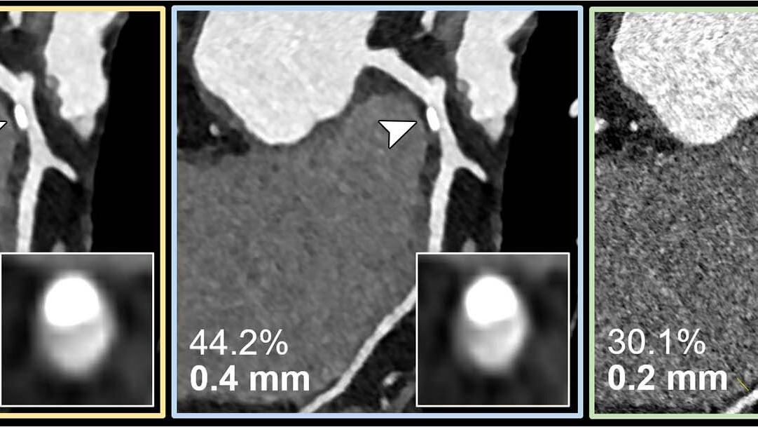
Emerging Technology in Cardiac Imaging
In the evolving world of cardiac imaging, a recent study published in Radiology, the esteemed journal of the Radiological Society of North America, illuminates a revolutionary technology: ultrahigh-spatial-resolution photon-counting detector computed tomography (PCD-CT). The study found that PCD-CT significantly improves the assessment of coronary artery disease (CAD), a leading cause of death worldwide.
PCD-CT, thanks to its enhanced image quality and superior spatial resolution compared to conventional CT, allowed for reclassification to a lower disease category in a remarkable 54% of patients in the study. This leap forward in technology could potentially lower healthcare costs and reduce unnecessary procedures by providing more accurate diagnostic information.
Addressing Limitations of Conventional Cardiac CT Angiography
The study further elaborates on the benefits of PCD-CT in addressing the limitations of conventional cardiac CT angiography. Notably, it reduces the overestimation of stenosis – a narrowing of the blood vessels – caused by calcium blooming. Calcium blooming is a common issue in patients with calcium buildup in the plaque of their coronary arteries, leading to an overestimation of the severity of the disease and therefore possibly unnecessary interventions.
PCD-CT demonstrated a reduction in overestimation of stenosis and a lower median degree of stenosis for calcified plaques compared to standard CT. However, the study notes that no substantial benefits were observed for mixed and non-calcified plaques, indicating that the technology may be most beneficial for patients with significant calcifications.
PCD-CT: A Potential Game-Changer in Patient Management
PCD-CT’s superior resolution and image quality could revolutionize patient management. By providing more accurate assessments of coronary artery disease, the technology has the potential to reduce unnecessary interventions and lower healthcare costs. This could lead to improved patient outcomes, more efficient use of resources, and better patient experiences.
Furthermore, the technology’s potential applications extend beyond CAD assessment. For instance, in preprocedural planning of transcatheter aortic valve replacement (TAVR), PCD-CT has shown promise. A study comparing ultrahigh resolution CT angiography (UHR CTA) with high pitch spiral CTA (HPS CTA) using a first generation dual source photon counting CT scanner found that UHR CTA demonstrated higher image quality compared to HPS CTA, providing reliable aortic annular assessments for TAVR planning.
Looking Ahead: The Need for Further Validation
Despite the promising results of PCD-CT in assessing coronary artery disease, the technology still requires further validation in real-world comparisons. Future research should focus on the potential impact of thin slice high tube voltage PCCT on overall image quality and radiation dose. Furthermore, as the technology continues to evolve, updated guidelines may be necessary for coronary CT angiography performed using PCCT.
In conclusion, PCD-CT represents an exciting development in the field of cardiac imaging. With its ability to provide high-quality images and reduce the overestimation of stenosis, it holds the potential to transform the diagnosis and management of coronary artery disease. However, as with any new technology, further validation is needed to fully understand its capabilities and limitations, and to ensure it is used effectively and safely in clinical practice.
