A Labrador retriever named Ava is back to running and playing with her family after her second double hip replacement, courtesy of Texas A&M University veterinarians, computerized tomography (CT)-guided planning, and 3D-printing technology.
When the two replacement hips Ava received as a puppy wore out in 2020, Texas A&M veterinarians were able to remove the old joints and replace them with new ones by using CT-guided planning, 3D-printed bone models and rehearsal surgeries to ensure the procedures would be successful.
From foster to family
Not many dogs go through four total hip replacements (THRs) in one lifetime, but Ava has always been special.
“Ava came to us at about 6 months old, back when we were dog foster parents living in Illinois,” said Ava’s owner, Janet Dieter. “After caring for more than 40 dogs, she was our first ‘foster failure’ that we ended up adopting. At the time, we had another black Labrador named Roscoe who was usually standoffish with the foster puppies, but he fell in love with Ava immediately and we knew she had to stay.”
Janet and her husband, Ken, always take the dogs under their care to obedience school, and Ava was no exception. It was there, however, that the couple began to notice something different about her.
“The subject of how to keep your dog from jumping on you came up and we realized that Ava never jumps on us,” Janet said. “We took her to our local veterinarian and they took an X-ray, which showed that Ava’s hips were basically out of their sockets.”
The Dieters were referred to an accomplished total hip replacement surgeon who performed THRs on Ava’s hips in 2013 and 2014.
“She was incredibly resilient,” Janet said. “She walked out of the hospital like nothing happened.”
For many years since then, Ava has been helping show the Dieters’ foster puppies the ropes by giving them someone to play with. When the Dieters moved from Illinois to Texas a few years ago, she took the change in stride.

In 2020, however, Ava faced new challenges when one of her replacement hips wore out.
“Over many years, the artificial ball had worn away the plastic liner protecting the metal wall of the artificial joint,” said Dr. Brian Saunders, a professor of small animal orthopedics and Small Animal Orthopedics Service chief at the Veterinary Medical Teaching Hospital. “The artificial ball then wore through the metal backing, causing a complete dislocation.”
While worn out THRs aren’t common in dogs, they can happen in joint replacements that have been in place for many years.
“When Ava’s original hips were placed, the liners in the replacement joints weren’t as advanced as they are today,” Saunders said. “Technology has improved now to where it’s less likely for that problem to happen. Complications like Ava’s aren’t terribly common, but when they do happen, they require advanced techniques to achieve a successful outcome.”
In addition to the dislocation, the erosion of the metal wall in Ava’s hip had caused tiny metal particles to build up around the joint and in her pelvic canal, forming a granuloma.
“A granuloma is basically a sack of soft tissue that’s trying to contain the metal debris,” Saunders said. “Ava had a large metal granuloma that was blocking access to the hip joint and affecting her internal organs. There was also a chance that it could cause her body to reject any THR revision implants.
“Metallosis—the erosion process that causes the metal debris to build up into a granuloma—can trigger cellular changes, leading to resorption or dissolution of bone around the new hip. It’s like putting the body in a defensive mode against outside objects,” he said.
Taking surgery to a new dimension
Because of the complexity of the surgery needed to remove the granuloma and fix Ava’s hip, the Dieters’ local veterinarian recommended that they visit Texas A&M’s orthopedic specialists.
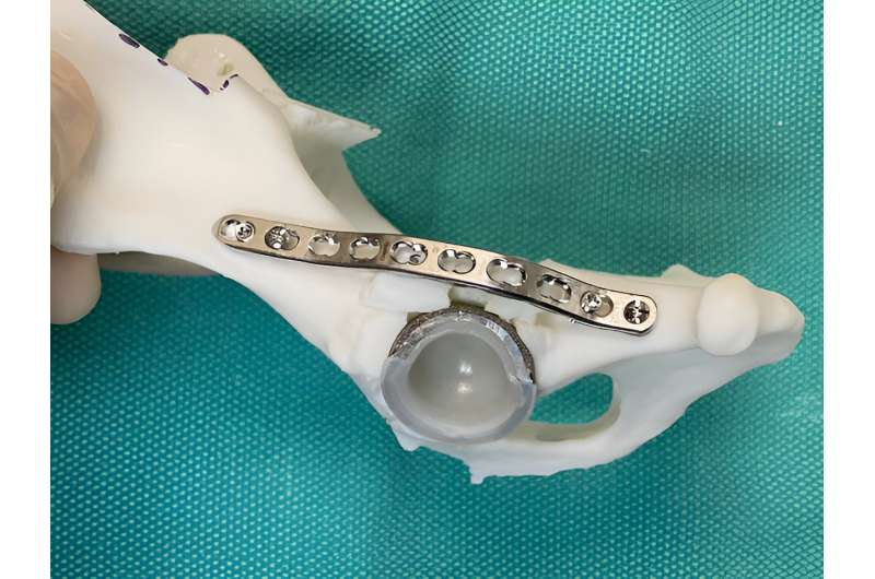
To make sure the complex surgery would be successful, Saunders used advanced CT-guided surgical planning and 3D-printing technology.

“We used computer-assisted 3D modeling to determine revision implant size and position,” Saunders said. “Basically, we printed a replica of Ava’s dislocated hip joint and planned exactly how to perform the revision operation using the 3D bone models. In fact, we sterilized the plastic models and used them in the operating room to help guide the revision surgery.”
Having 3D-printing technology right inside the VMTH is a huge advantage for surgical teams.
“If you don’t have your own 3D-printing program, you have to send a CT scan to a third-party company using a fee-for-service process. This can be challenging in regard to turnaround time and you often lose the ability to be involved in the planning process,” Saunders said.
Having a replica of Ava’s hip was especially helpful considering that she had a granuloma complicating things.
“To avoid a THR rejection, we used the CT scan and worked with the Soft Tissue Surgery group to remove as much of the metal granuloma from the pelvic canal before we came back and performed the THR revision. Then, when we did the revision, we were able to finish removing the remainder of the granuloma from the other side,” Saunders said. “Planning using 3D models and collaborating with the Soft Tissue team were two huge contributing factors to our success.”
Even though Ava’s first hip revision went well, her challenges weren’t over just yet. A few weeks after the first surgery, Ava’s other THR liner also wore out and dislocated. She had to return to the VMTH for a second hip revision.
“Thankfully, the second hip wasn’t quite as affected as her first and we already had her 3D bone models from the recent surgery, so the second hip revision surgery was more straightforward,” Saunders said.
Ava, who is now 12 years old, hasn’t let her hip history slow her down.
“She still zips all over the backyard and through our exercise course,” Janet said. “She will even jump over the couch.”
“When she started showing signs of her first hip wearing out, we thought it might mean the end, and we were devastated,” Ken said. “But the veterinarians at Texas A&M were able to give her life again.”
Provided by
Texas A&M University
Citation:
Veterinarians use 3D printing technology to assist in double hip replacement surgery for a dog (2023, November 16)
retrieved 16 November 2023
from https://phys.org/news/2023-11-veterinarians-3d-technology-hip-surgery.html
This document is subject to copyright. Apart from any fair dealing for the purpose of private study or research, no
part may be reproduced without the written permission. The content is provided for information purposes only.
